Initial Situation

Cell growth is monitored in biology, biotechnology, and pharmaceuticals for numerous applications, such as testing different substances. However, manual cell counting in microscopy images is time-consuming. Automated image analysis can take over this step. The challenge: typical lab PCs offer limited computing power, yet fast and reliable results are needed.

Markierung Algorithmus Zellen

Project Overview

Client
Manufacturer of zenCELL owl

Sector
Medical & Life Science

Solution
Deep-learning cell counting directly on the lab PC.

System Environment
zenCELL owl, compact and stackable microscopy system for incubators, PC-based analysis

Special Feature
optimized for local operation on resource-constrained lab PCs

Application
simultaneous monitoring of 24 samples

SCS’s Contribution

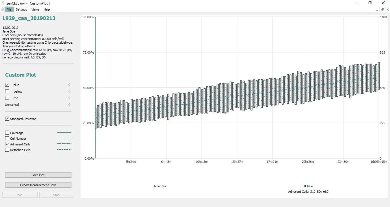
SCS developed a deep-learning algorithm for automatic cell counting and specifically optimized the neural network for fast analyses on limited computing power.
CoverageOverTime

Result

With the algorithm developed by SCS, cell counts can be determined automatically, even on existing lab hardware. The zenCELL owl can simultaneously monitor up to 24 samples in the incubator and display their development on a PC, with the software automatically determining both cell count and confluence. This eliminates a time-consuming manual step for scientists. According to the project assessment, automatic analysis saves many hours with comparable quality, without the need to purchase more powerful hardware.

24 samples

monitored simultaneously in the incubator.

many hours

less manual effort with comparable quality.

no new hardware

optimized for existing lab computing power.

Services Used

Algorithms, ML & AI

Computer Vision

Edge Computing

Medical & Life Science

Smiling man with dark hair and a mustache in a white shirt; 'SCS Contact' is blurred against a colorful background.

Your Contact Person

Beat Hörmann

Healthtech & Innovation
+41 43 456 16 00

For questions about Machine Learning, Computer Vision, and AI models for resource-constrained hardware.

More Case Studies

  • Microscopic image of tissue with a scatter plot, overlaid data fields, and medical technology software.

    ariadne.ai

    Web Viewer for Microscopy Images

    High-performance Web Viewer for very large image data.

  • A black-and-white OCT scan using the "Medical Technology" software clearly shows the layers of the retinal structures.

    Visual Innovation Through AI in Ophthalmology

    AI segmentation of medical structures in OCT scans.

  • A white and red high-speed train glides environmentally friendly through the green landscape under a blue sky.

    SBB

    Trustworthy AI for the Measuring Wheelset

    Explainable AI for Force Measurement on Wheel and Rail.