Initial Situation

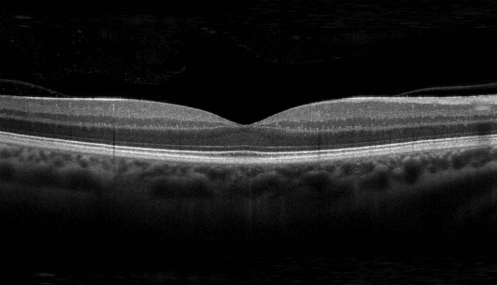
OCT scans allow for the non-invasive recording of the retina’s fine layer structure. OCT is frequently used in clinics to diagnose major eye diseases, such as age-related macular degeneration, glaucoma, or diabetic retinopathy. However, the manual evaluation of three-dimensional image data is time-consuming and requires medical expertise. AI-supported image analysis can help to automatically segment relevant structures and evaluate large quantities of OCT data in a reproducible manner.
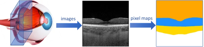
Diagramm: Auge, Netzhautscan und beschriftete Pixelkarte, nacheinander mit Pfeilen dargestellt.

Project Overview

Field of Application
Ophthalmology and Research

Area
Medical & Life Science

Solution
AI segmentation of medical structures in OCT scans.

System Environment
On-premise platform, dockerized containers, web interface for anonymized OCT scans

Key Features
Cross-device OCT segmentation, synthetic tumor images with GANs, reproducible AI development

Evidence
Results on OCT segmentation published scientifically (PLOS ONE, 2019)

The Contribution of SCS

SCS developed a modified U-Net CNN for the automated segmentation of OCT images and set up a reproducible development environment for training, evaluation, and operation. For rare tumor images, SCS additionally investigated methods for synthetic data generation.

Drei Augen-Scans, auf denen verschiedene erhabene Bereiche in der mittleren Netzhaut durch rote Kreise hervorgehoben sind.

Result

In the scientific evaluation, the automated segmentation of the examined eye structures was within the variability of human evaluations. The model was deployed as a dockerized service on a local platform. Anonymized OCT scans can be evaluated via a web interface, and the areas and volumes of the segmented eye structures can be determined. The approach supports different OCT recording methods and devices. Scientifically published: “Validation of automated artificial intelligence segmentation of optical coherence tomography images”, PLOS ONE, 2019.

At a Human Level

Segmentation of eye structures within the range of manual evaluations.

Cross-Device

The model works for various OCT devices.

PLOS ONE 2019

Scientifically published evaluation of OCT segmentation.

Services Provided

Algorithms, ML & AI

Computer Vision

Medical & Life Science

Smiling man with dark hair and a mustache in a white shirt; 'SCS Contact' is blurred against a colorful background.

Your Contact Person

Beat Hörmann

Healthtech & Innovation
+41 43 456 16 00

For questions regarding machine learning, medical image segmentation, and reproducible AI development.

Further Case Studies

  • White lab device labeled "zenCELL owl" utilizing edge computing; blurred lab utensils in the background.

    Manufacturer of zenCELL owl

    AI at the Edge – Counting Cells

    Deep-learning cell counting directly on the lab PC.

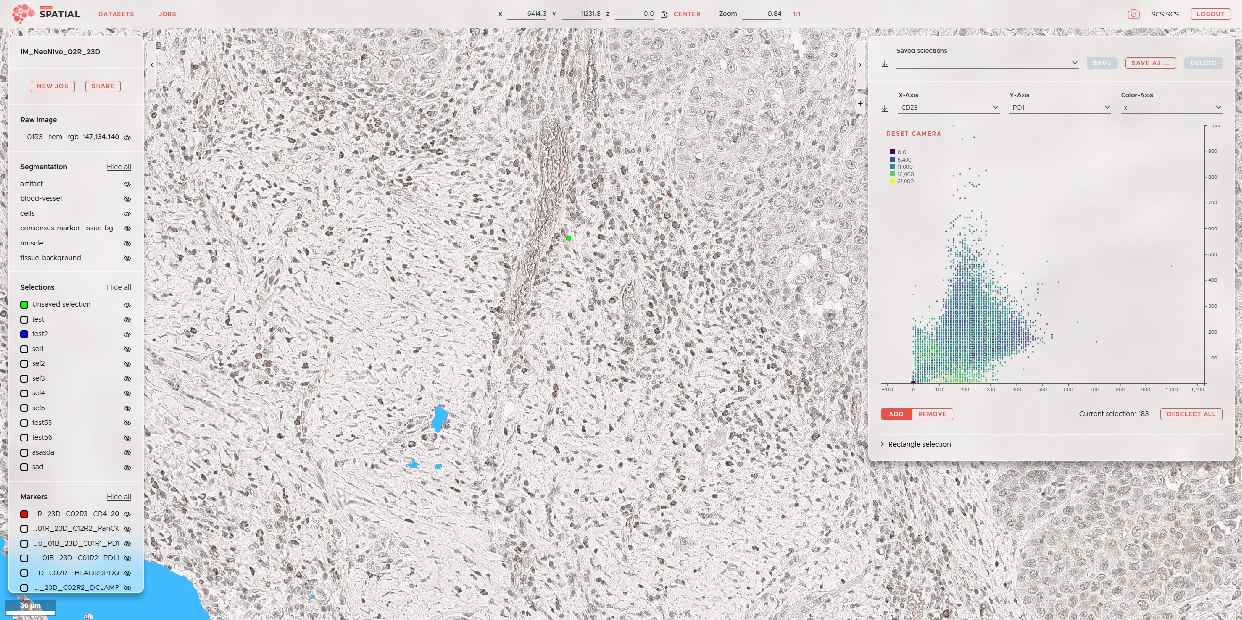
  • Microscopic image of tissue with a scatter plot, overlaid data fields, and medical technology software.

    ariadne.ai

    Web Viewer for Microscopy Images

    High-performance Web Viewer for very large image data.

  • A white and red high-speed train glides environmentally friendly through the green landscape under a blue sky.

    SBB

    Trustworthy AI for the Measuring Wheelset

    Explainable AI for Force Measurement on Wheel and Rail.