Initial Situation

Automated analysis of biomedical images quickly generates several terabytes of data. Microscopy images are available in various resolutions and image planes and are supplemented by segmentations, annotations, and analysis data. Making these data volumes quickly, fluidly, and interactively accessible in the browser places high demands on the architecture and performance of the frontend.
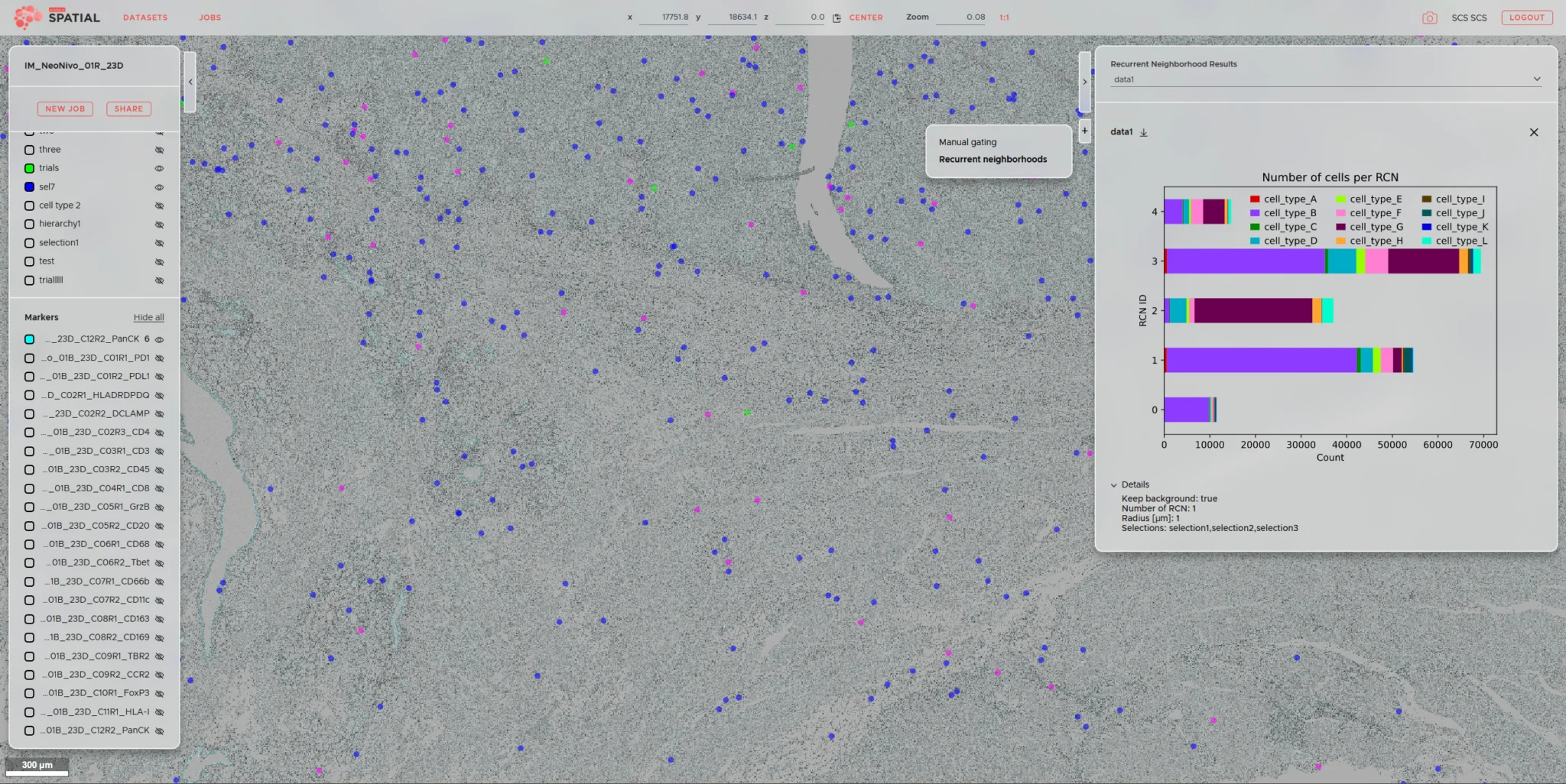
Map with grouped blue and pink dots, a bar chart, and an image processing analysis for cell counting.

Project Overview

Client
ariadne.ai

Sector
Medical & Life Science

Solution
High-performance Web Viewer for very large image data.

System Environment
Neuroglancer, Angular, D3.js, containerized deployment

Scope
Complete frontend development from requirements analysis and wireframes through UI and UX to implementation, deployment, and performance optimization

Data Volumes
Image data in the terabyte range, multiple resolution levels and image planes

SCS’s Contribution

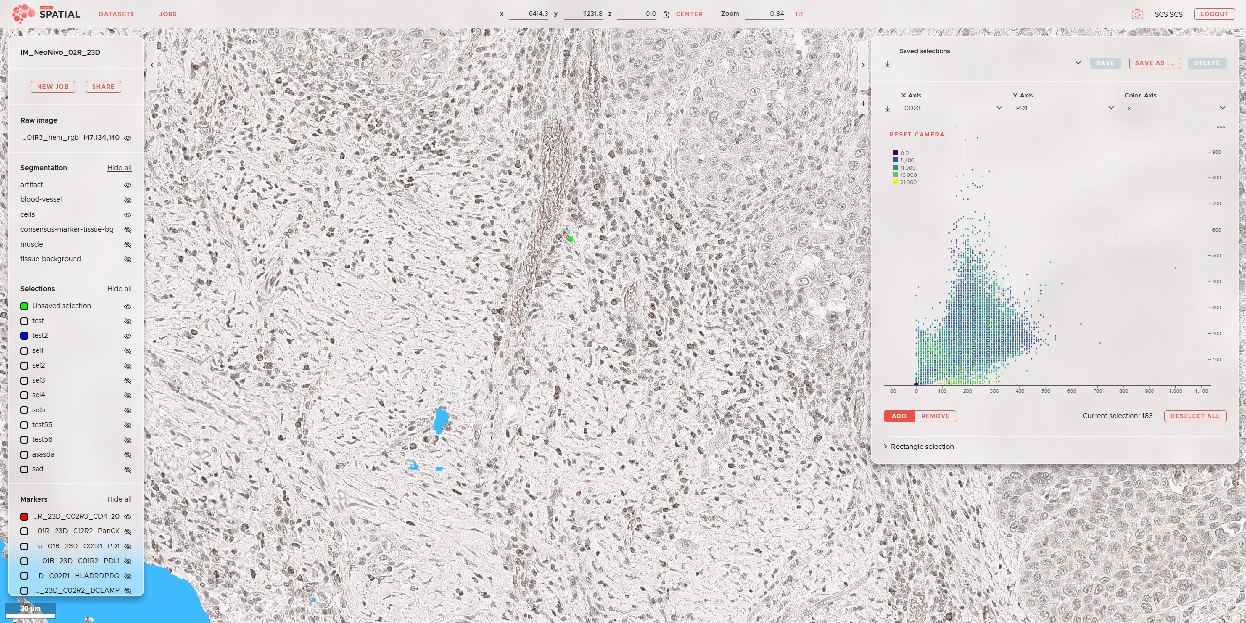
SCS developed the complete frontend for Ariadne SPATIAL for ariadne.ai. The web viewer interactively displays large image data, integrates ariadne.ai’s processing results, and allows users to select data areas, segmentations, and analysis parameters directly in the browser.

Ariadne Bild 4

Result

With Ariadne SPATIAL, even very large microscopy datasets can be interactively explored in the browser. Users can switch between resolution levels and image planes, select segmentations, and link analysis data directly with the underlying images. SCS was responsible for the entire frontend, from requirements analysis and wireframes through UI and UX to implementation, containerization, and performance analysis and optimization.

several terabytes

Image data interactively displayed in the web viewer.

complete frontend

from requirements analysis and UX to performance optimization.

multiple data layers

Images, segmentations, annotations, and metadata visualized together.

Services Used

High Level Software

UX / UI

Data Visualization

Medical & Life Science

Smiling man with dark hair and a mustache in a white shirt; 'SCS Contact' is blurred against a colorful background.

Your Contact Person

Beat Hörmann

Healthtech & Innovation
+41 43 456 16 00

For questions regarding machine learning, medical image segmentation, and reproducible AI development.

Further Case Studies

  • White lab device labeled "zenCELL owl" utilizing edge computing; blurred lab utensils in the background.

    Manufacturer of zenCELL owl

    AI at the Edge – Counting Cells

    Deep-learning cell counting directly on the lab PC.

  • A black-and-white OCT scan using the "Medical Technology" software clearly shows the layers of the retinal structures.

    Visual Innovation Through AI in Ophthalmology

    AI segmentation of medical structures in OCT scans.

  • A boy blows into a medical device, strengthening his digital resilience, while an adult watches him in a bright room.

    ndd Medizintechnik AG

    Cloud Platform for Medical Measurement Data

    Cloud integration of existing medical devices with patient records.