Background

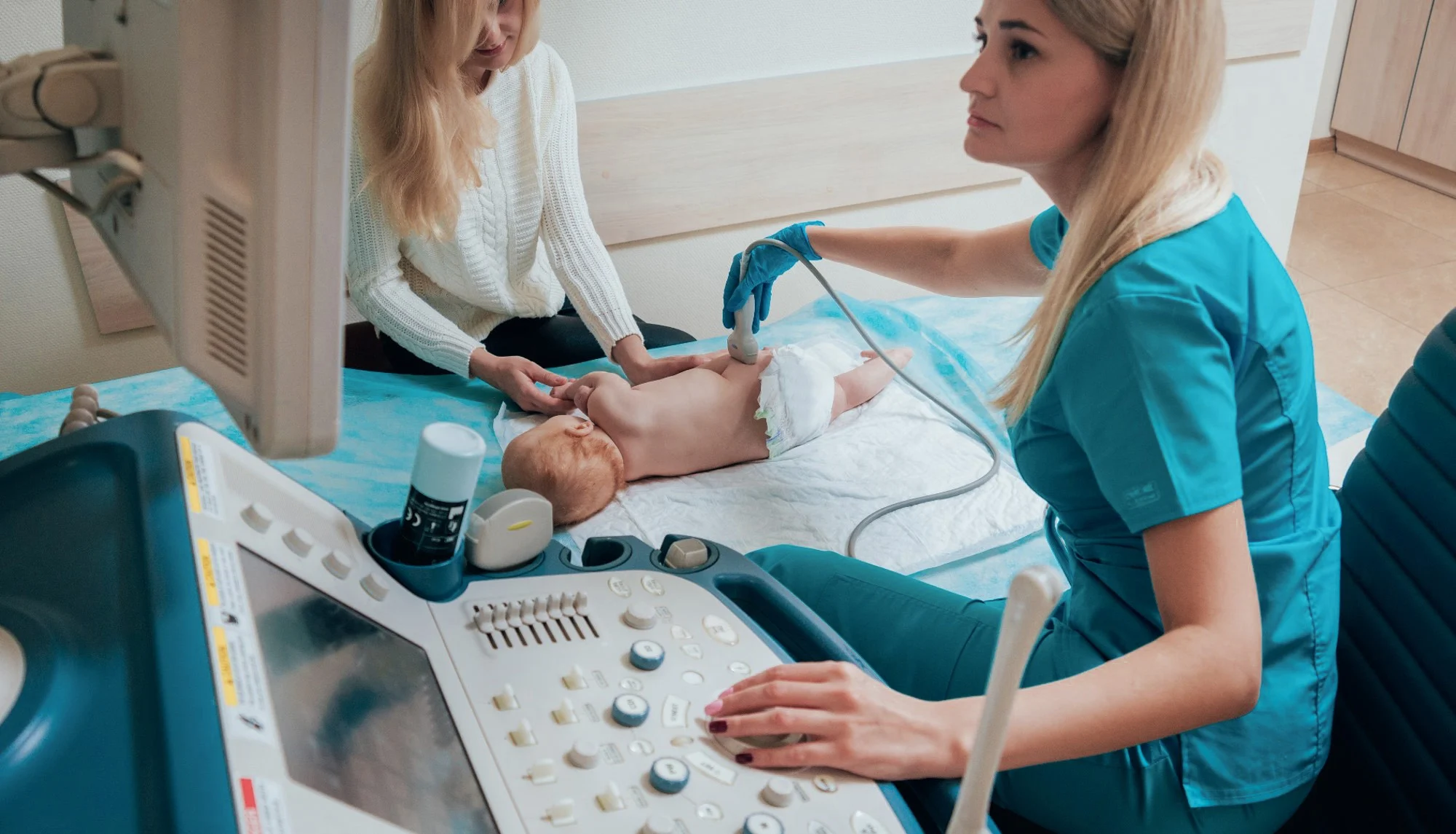
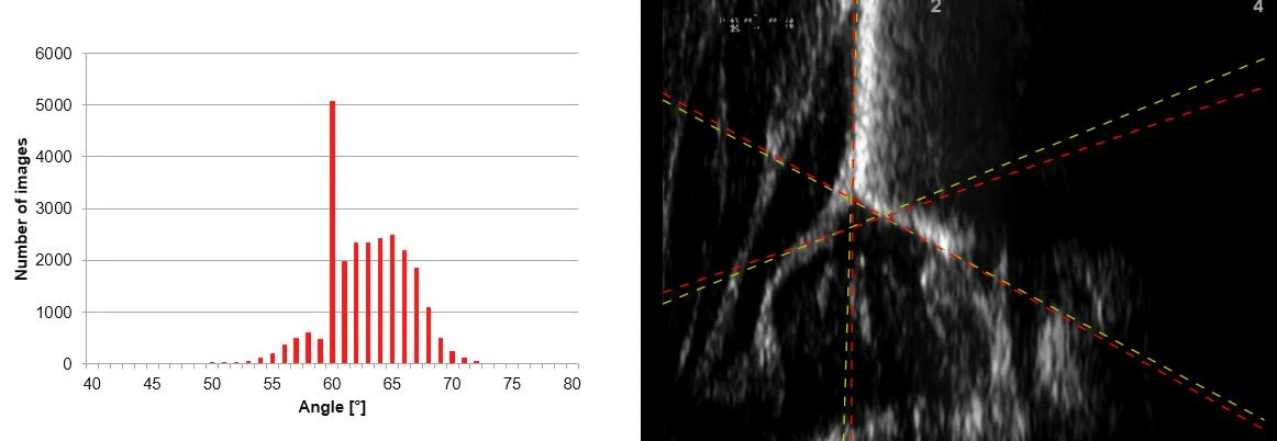
Approximately 3 percent of all newborns are born with hip malalignment. If detected early via ultrasound, it can usually be treated easily, for example with an abduction brace for a few weeks. If it remains undetected, this hip dysplasia leads to hip surgery before the age of 60 in 30 percent of those affected. The Graf alpha angle is particularly decisive for the assessment. However, its determination depends on the placement of the measurement lines and can vary between evaluators. The existing clinical data also showed a noticeable clustering of measured values exactly at the threshold of 60 degrees.
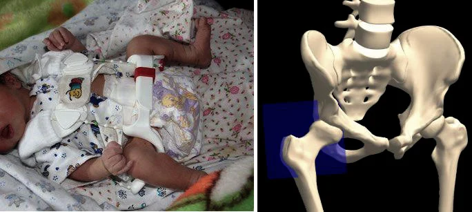
Säugling mit einer Hüftorthese und einem 3D-Modell eines Hüftgelenks und eines Beckens.

Project Overview

Field of application
Pediatrics, ultrasound diagnostics of the infant hip

Area
Medical & Life Science

Solution
AI-supported assessment of infant hips in ultrasound.

System environment
Ultrasound images of the infant hip, Graf alpha and beta angles

Partners
Dr. Stefan Essig, Dr. Thomas Baumann (Pediatricians)

Evidence
Joint study, scientifically published in Ultraschall in der Medizin

The SCS Contribution

SCS trained various algorithms using ultrasound images to determine the hip angle objectively and reproducibly, and proved the approach in a joint study with specialists and pediatricians.
Balkendiagramm der Bildwinkel und Graustufenabtastung mit farbigen gestrichelten Linien, die sich auf der rechten Seite kreuzen.

Result

The comparison with independent data from pediatricians shows that the algorithm determines the hip angle reliably. The joint study confirmed that the angle can be determined objectively and reproducibly using deep learning. More precise and reproducible angle determination can support early diagnosis and alert physicians to unsuitable recordings at an early stage.

approx. 3 percent

of newborns are born with hip malalignment.

30 percent

of undetected cases lead to hip surgery before the age of 60.

scientifically published

Study results in Ultraschall in der Medizin.

Services Utilized

Algorithms, ML & AI

Computer Vision

Medical & Life Science

Smiling man with dark hair and a mustache in a white shirt; 'SCS Contact' is blurred against a colorful background.

Your Contact Person

Beat Hörmann

Healthtech & Innovation
+41 43 456 16 00

For questions regarding machine learning, medical image analysis, and objective, reproducible diagnostic support.

Further Case Studies

  • White lab device labeled "zenCELL owl" utilizing edge computing; blurred lab utensils in the background.

    Manufacturer of zenCELL owl

    AI at the Edge – Counting Cells

    Deep-learning cell counting directly on the lab PC.

  • Microscopic image of tissue with a scatter plot, overlaid data fields, and medical technology software.

    ariadne.ai

    Web Viewer for Microscopy Images

    High-performance Web Viewer for very large image data.

  • A black-and-white OCT scan using the "Medical Technology" software clearly shows the layers of the retinal structures.

    Visual Innovation Through AI in Ophthalmology

    AI segmentation of medical structures in OCT scans.